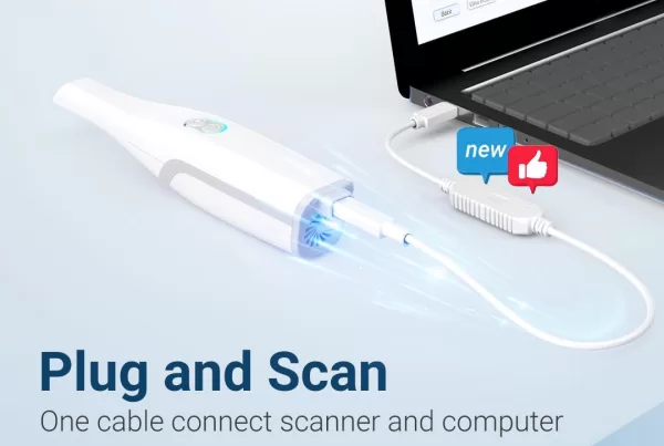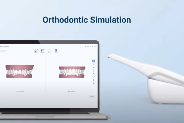
It is quite familiar to those who work as a dentist about intraoral impression. In recent decades, another word has become more and more well-known—IOS(intraoral scanning). The last decade has seen an increasing number of optical intraoral scanning devices (Intraoral Scanner INO100, etc.). And they are based on different techniques.
Light Projection and Capture
There is a clear difference between passive and active techniques used in intraoral scanning within the 3D reconstruction field.
Passive techniques
Passive techniques only use ambient lighting to enlighten intraoral tissues, which are highly dependent on the object’s texture.
Active techniques
For active techniques, however, the camera projects white, red or blue structure lights onto the surface of the object. It is less reliant on the real texture and color of tissues for reconstruction.
Points emission
In this way, a luminous point is projected onto an object and the distance is calculated by triangulation.
Network emission
This means light pattern projection. A video can take several images per second in a continuous data flow and then reconstruct the object’s surface.
Distance to Object Technologies
Active Wavefront Sampling (AWS) is a 3D surface imaging technique that employs only one camera and an AWS module. The AWS module is an off-axis aperture, which moves on a circular path around the optical axis. Theoretically, AWS imaging allows any system with a digital camera to function in 3D.
Triangulation
As can be seen from the picture, distance BC could be determined according to the formula
BC = AC × sin ̂A/sin ̂A+̂C

Confocal Imaging
Confocal imaging is an IOS technique that involves the acquisition of focused and defocused images from specific depths. This technology can detect the sharpness area of the image, so to conclude distance to the object. And the distance is determined according to the focal distance.
Stereophotogrammetry
Stereophotogrammetry is the process of estimating the 3D coordinates of points on an object. We can achieve that by using measurements taken in two or more photographic images obtained from different perspectives. Therefore, the image is calculated from a collection of points obtained along an x, y, and z coordinate system.
3D Reconstruction Technologies
3D reconstruction technology refers to the process of converting multiple 2D medical image slices to a 3D anatomical model. It enables the accurate display of the anatomical structure and lesion’s spatial position, size, geometric shape, as well as its spatial relationship with the surrounding tissues.

